Figure 5.
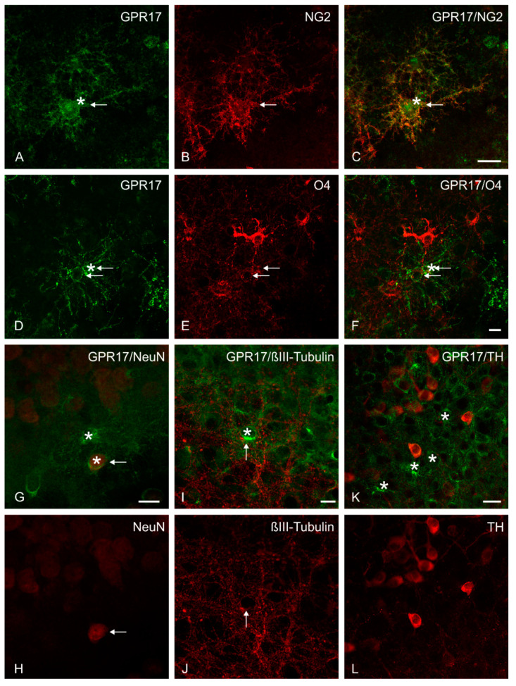
Multiple immunofluorescence study. Representative confocal images of GPR17 expression in organotypic slice co-cultures. At DIV 10 an intense GPR17 immunoreactivity was observed on NG2-positive cells (A–C) and on O4-positive cells (D–F). A low expression of GPR17 (stars) on a small number of cells was observed on (G,H) NeuN-positive cells and (I,J) βIII-Tubulin-positive neurons (the thin arrows indicate the co-expression). No co-localization, but a number of GPR17-positive cells (stars) in the proximity were found on (K,L) TH-positive cells. Scale bars: (A–C) = 20 µm; (D–J) = 10 µm; (K,L) = 20 µm.

