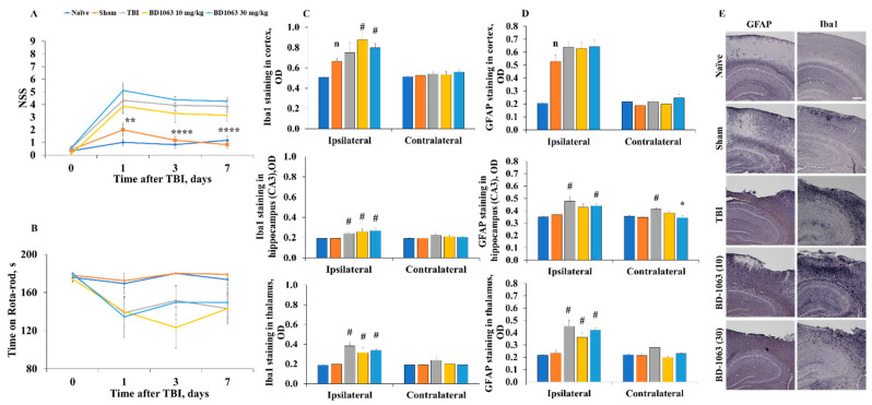
Figure 4.
Posttraumatic behavioral and histological outcomes after pretreatment with the S1R antagonist BD-1063. (A) TBI mice had a significantly higher NSS up to 7 days postinjury. (B) All experimental groups spent similar time on the RR. BD-1063 treatment did not influence behavioral outcomes after TBI. Data are presented as the means ± SEM (naïve n = 6, sham n = 9, TBI n = 15, BD-1063 10 mg/kg n = 7, BD-1063 30 mg/kg n = 8). p values for differences between groups were calculated using RM two-way ANOVA followed by Fisher’s LSD test: * p < 0.05, ** p < 0.01, **** p < 0.0001 sham vs. TB. OD of the intensity of (C) Iba1 and (D) GFAP staining in the cortex, hippocampus, and thalamus. TBI induced significantly increased staining for Iba1 in the ipsilateral thalamus and for GFAP in the ipsilateral hippocampus and thalamus. (E) Representative images of Iba1 and GFAP staining at 7 days postinjury (total magnification = 40×, scale bar = 100 µm). Data are presented as the means ± SEM (n = 5). p values for differences between groups were calculated using the Kruskal–Wallis test followed by Dunn’s test: # p < 0.05 compared with the sham group, * p < 0.05 compared with the TBI group. Differences between naïve and sham groups were calculated using the Mann–Whitney U-test: n p < 0.05 compared with the naïve animals.

