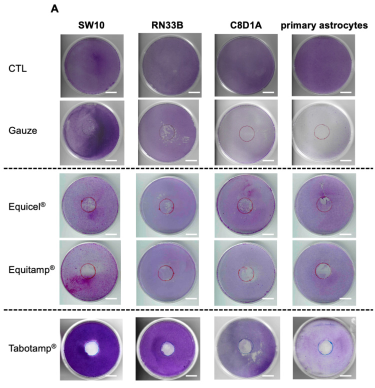
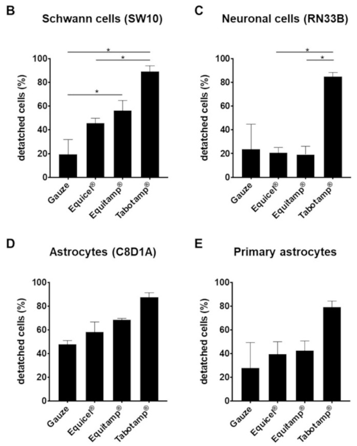
Figure 2.
Detachment of cell monolayer after incubation with Equicel® and Equitamp®. Exemplary crystal violet staining of each ORC and cell line or primary cells. The results of untreated cells (control, CTL) and after incubation with gauze and Tabotamp® have already been published and are shown for comparison [10]. Cells were grown to complete confluence in 60 cm2 cell culture dishes and incubated with Equicel® or Equitamp®. Bar = 2 cm (A). In addition, the means and SEM of quantified cell detachment analysis of three independent stained dishes from Schwann cells (B), neuronal cells (C), immortalized astrocytes (D) and primary astrocytic cultures (E) are shown (* p < 0.05).


