Figure 1.
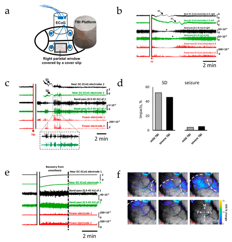
Spreading depolarization is the earliest and most common electrophysiological event after TBI. The upper two traces in each panel show raw ECoG recordings (band-pass: 0.02–100 Hz). The 3rd and 4th traces show band-passed (0.5–45 Hz) activity. Recordings from both the right (black) and left (green) hemispheres are shown. The 5th and 6th traces (red) show the squared activity (power near DC-ECoG). (a) Schematic representation of the general experimental setup. (b) Recording showing TBI-induced SDs recorded from both hemispheres. (c) Recording showing TBI-induced spreading depolarization with seizure activity immediately before and after SD; post-SD seizure is shown with expanded timescale. (d) Occurrence rates of SDs and seizures following mild and severe TBI. (e) Recording from a non-injured control; recovery from anesthesia is associated with a high-amplitude activity, which returns to pre-impact activity after regain of locomotion (dotted line). (f) Intravital microscopy showing changes in intrinsic optical signals during SD; changes in IOS are superimposed onto brain images; SDs propagated medially toward the midline; the dotted line represents the SD fronts. A: anterior; P: posterior; L: lateral; M: medial.

