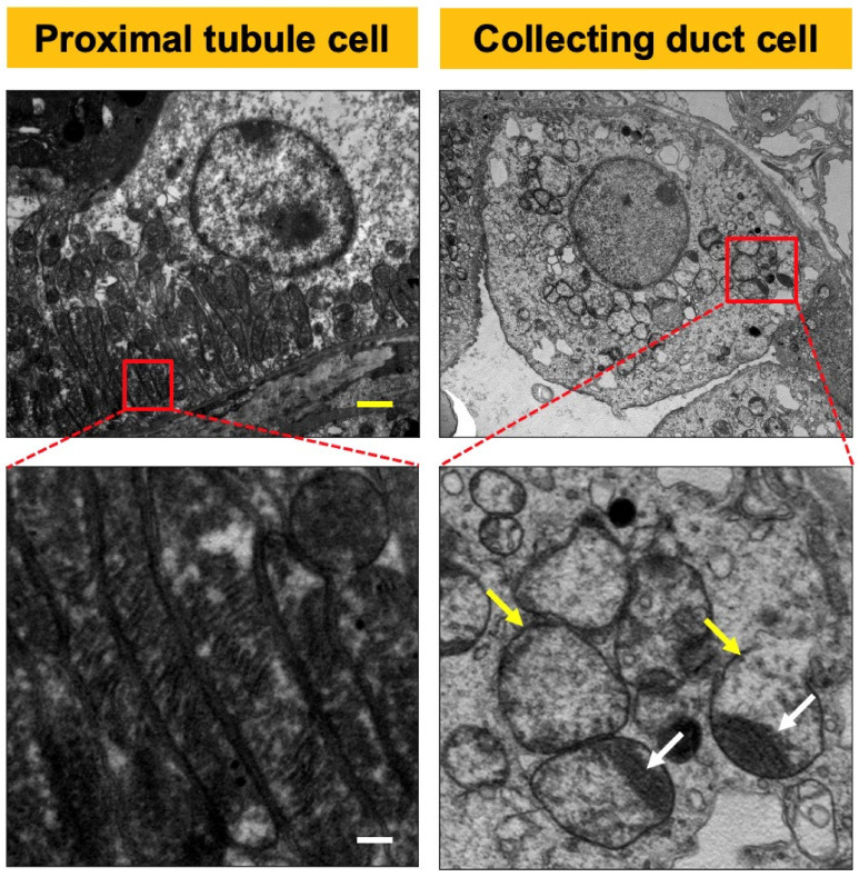
Figure 2.
Photos of electron microscopy of CR6-interacting factor-1 knockout (CRIF1-KO) sham mouse kidney. The mitochondria of collecting duct cells were swollen (white arrow) and showed the destruction of cristae (yellow arrow) in the kidneys of CRIF1-KO mice. The mitochondria of proximal tubular cells showed normal morphology in CRIF1-KO mice. Yellow scale bar, 2 μm. White scale bar, 0.2 μm.

