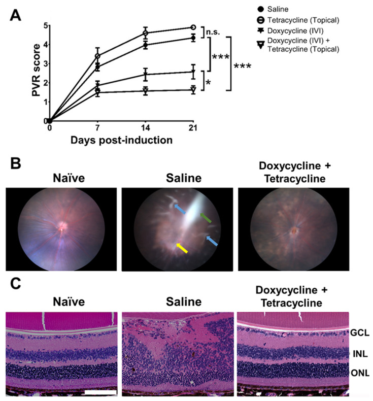
Figure 3.
Doxycycline and tetracycline treatment reduces PVR in mice. (A) After PVR induction and treatment with saline and doxycycline by IVI injection or tetracycline topically, mice were monitored for disease scores in the fundus using a slit lamp on the indicated days post-induction. The data represent means ± SEM (error bars) of ≥10 mice per data point. Note: *, p < 0.05 and ***, p < 0.001 via the one-way ANOVA followed by the Holm–Sidak test on day 21 post-induction. Note: n.s.: not significant. (B) The representative fundus images of naïve mice or mice with PVR induction and treated with saline or doxycycline and tetracycline with ERM (green arrow), inflammation (yellow arrow), and retinal detachment (blue arrows) are shown. (C) The eyes of mice described in (B) were harvested 21 days after induction, sectioned, and stained with H&E. GCL: ganglion cell layer; INL: inner nuclear layer; ONL: outer nuclear layer. Scale bar, 100 μm. Data are representative of at least 2 experiments.

