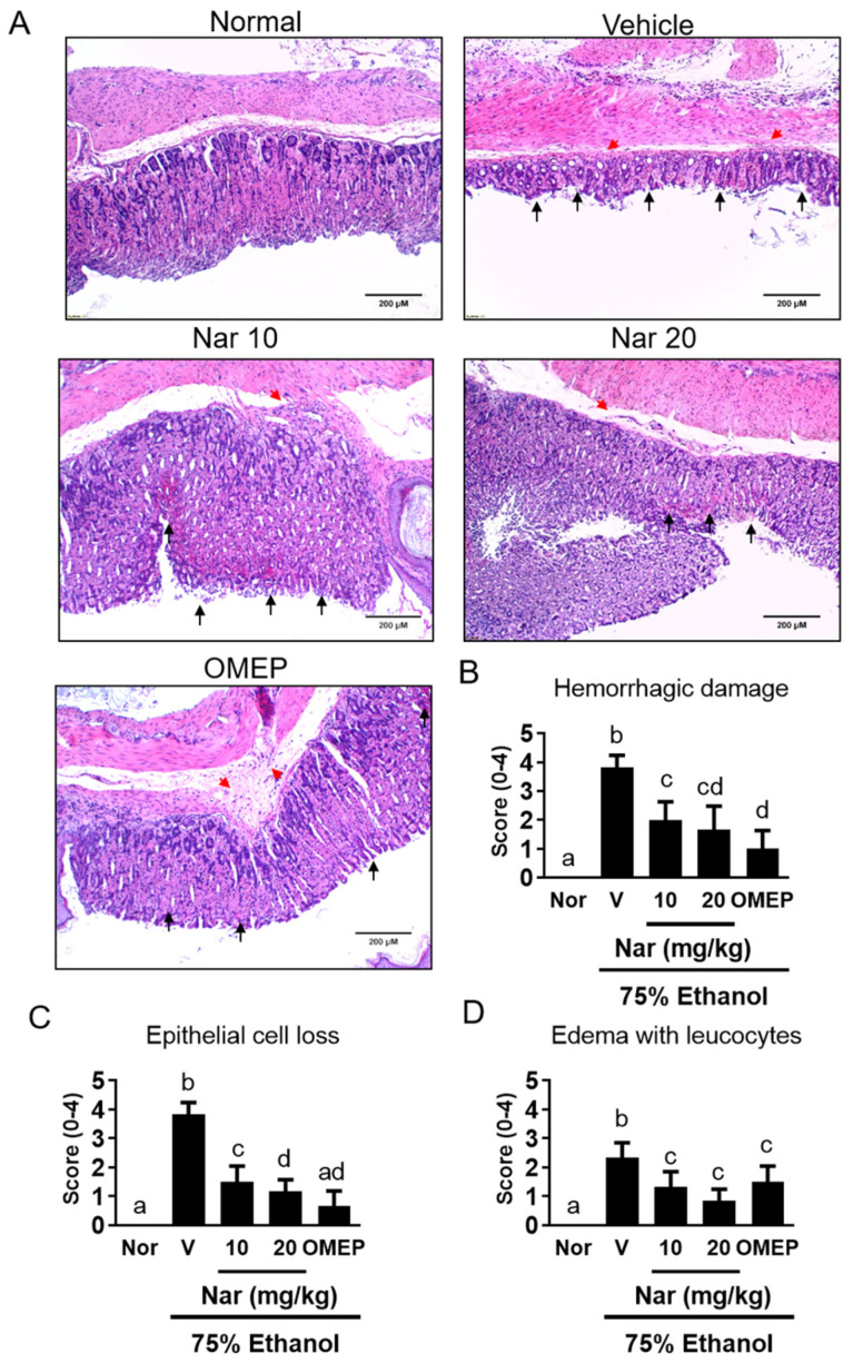
Figure 2.
(A) Histological assessment of the effects of naringenin on acute gastric mucosal injuries in ethanol-induced mice. The gastric tissue (n = 6/group) was fixed with 4% paraformaldehyde and sectioned for HE staining (magnification ×100). Red arrowheads indicated the presence of edema, hemorrhagic damage. Black arrowheads indicated the loss of epithelial cells.. The scores for (B) hemorrhagic damage, (C) epithelial cell loss and, (D) edema with leucocytes. The vehicle (10% dimethyl sulfoxide (DMSO) and 90% glyceryl trioctanoate), naringenin (Nar, 10 and 20 mg/kg) and omeprazole (OMEP, 20 mg/kg; used as a positive control) were given orally for a period of 3 days. After fasting for 12 h prior to the experiment, mice were fed orally with ethanol (0.5 mL/100 g body weight) to induce the acute ulcer. After 4 h, mice were sacrificed. The data obtained from individual animal samples per group were averaged (n = 6); values represent mean ± standard deviation (SD). Statistical comparison was analyzed by a one-way ANOVA, followed by Tukey’s multiple comparison tests. Bars not sharing a common letter represent a statistically significant difference from each other (p < 0.05). In the normal control group, the mice only received vehicle (Normal). Nor, normal; V, vehicle.

