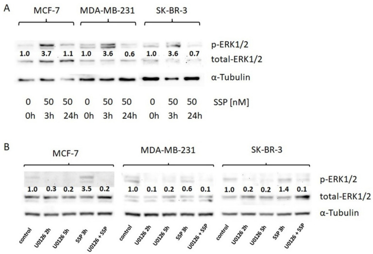
Figure 4.
Western blot analysis of ERK1/2 activation in breast carcinoma cells. (A) Breast carcinoma cells were cultured in DMEM, 10% FCS, and directly solubilised (0 h) or solubilised after incubation with 50 nM of SSP for the indicated time spans. (B) Breast carcinoma cells were directly solubilised (control) or, before solubilisation, treated for the indicated time spans with the MEK inhibitor U0126 (20 μM) or for 3 h with 50 nM of SSP either in the absence or presence of 20 μM of U0126. α-Tubulin was used as a loading control. Numbers show fold change compared to controls (set as “1.0”).

