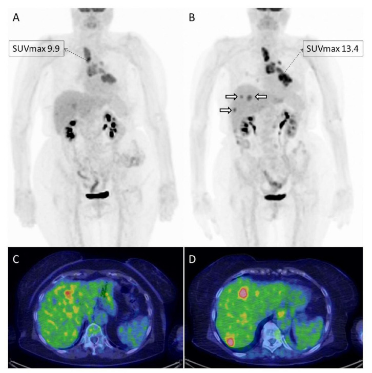
Figure 2.
Example of a 78-year old female with advanced NSCLC treated with nivolumab and imaged with [18F]FDG PET/CT at baseline (A,C) and after 4 cycles of therapy (B,D). The patient resulted in overall stable on morphological imaging performed prior to PET/CT, which on the contrary documented a progressive metabolic disease. In fact, the tumor had an increase in metabolism (SUVmax and MTV), and showed the appearance of new lesions in the liver ((B); white hollowed arrows), only partially detectable on baseline imaging.

