
Figure 3.
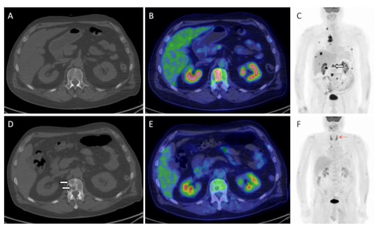
Herein, the imaging findings of a 66-year old male with metastatic NSCLC investigated before (A–C) and after 3 cycles of pembrolizumab (D–F). An overall response to treatment is easily visible on MIP (maximal intensity projection) images (C,F), including a complete metabolic remission of all bony lesions ((C); white hollowed arrow). On the contrary, morphological imaging proved the appearance of a new bone lesion in the first lumbar vertebra ((A,D); white arrows), which in fact corresponded to a healed metastasis on PET/CT (B,E). Note also the appearance of diffuse thyroid uptake ((F); red arrow), consistent with thyroiditis, one of the irAEs that typically predicts treatment response and good patient’s outcome.