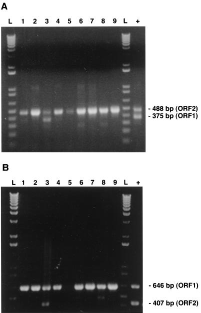
FIG. 5.
Detection and typing of PCV in a panel of positive clinical specimens (lung tissue; lanes 1 to 9) by both mPCR methods. As described in the Materials and Methods section, in both mPCR methods, two sets of primers were used simultaneously with DNA extracted from lungs of the affected pigs to amplify fragments of the ORF1 and ORF2 genes of PCV-1 but only the ORF1 or ORF2 fragment of PCV-2. (A and B) For only one of the clinical samples tested, an mPCR profile of a PCV-1 strain was observed. As expected, by the mPCR1 method (A), a fragment of 488 bp specific for the ORF2 genes of strains of both genotypes could be amplified for all positive samples, whereas for only one sample (lane 3), the ORF1 fragment (375 bp) could be amplified. By the mPCR2 method (B), a fragment of 646 bp specific for the ORF1 genes of both genotypes could be amplified for all positive samples, whereas for only the same sample described in panel A, the 407-bp fragment specific for the ORF1 of PCV-1 could be amplified (lane 3). Lane L, 1-kb DNA ladder; lane +, DNA extracted from purified PCV-1.

