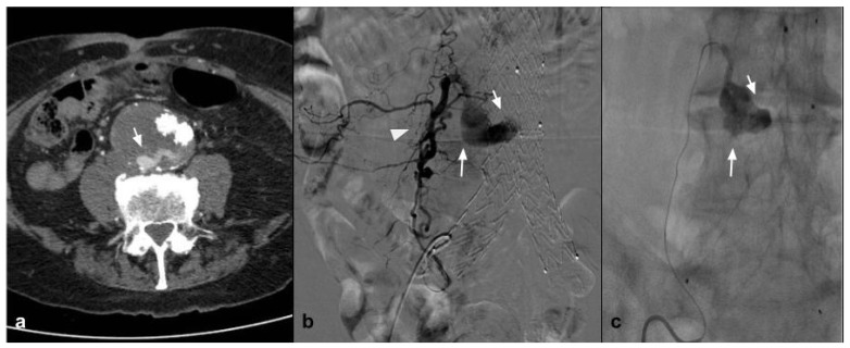
Figure 3.
(a) Axial CTA shows, the presence of Type II endoleak after EVAR, supplied by lumbar arteries (white arrow). (b) DSA performed with microcatheter positioned in a lumbar branch through the ilio-lumbar artery highlights the presence of hypertrophic lumbar circles (white arrow head) with sac refuelling (white arrow). (c) Post-procedure DSA shows the cast of Squid 12, which completely occupies the space of the endoleak in the sac (white arrow).

