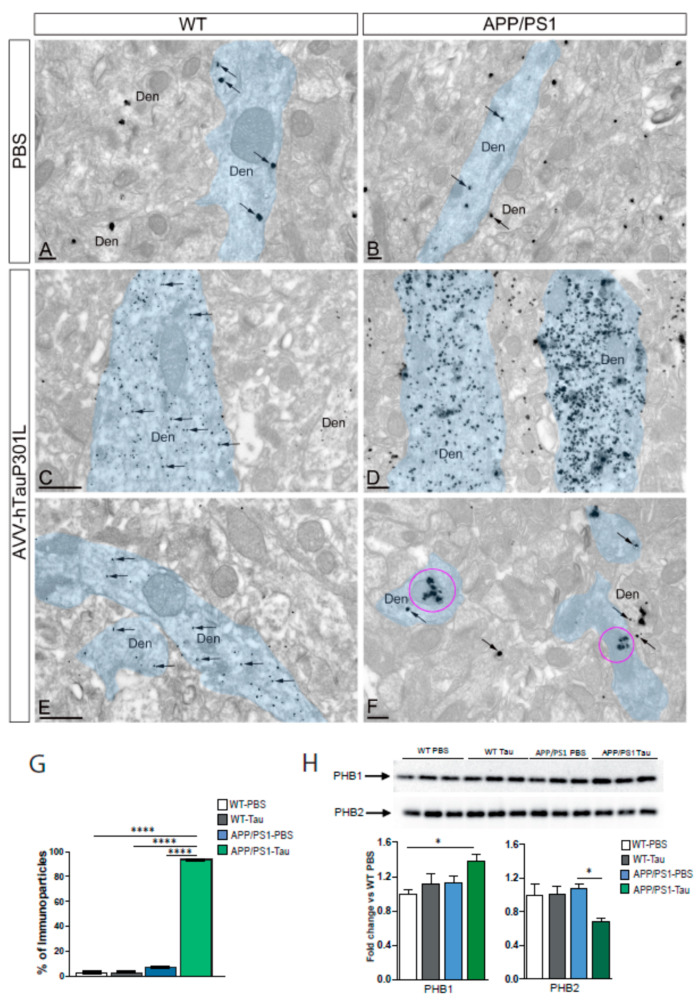
Figure 5.
Electron micrographs showing the distribution of immunoparticles for tau in the CA1 region of mice hippocampi using a pre-embedding immunogold technique. (A,B) In WT and APP/PS1 in control group (PBS), few immunoparticles for tau (arrows) were observed in the cytoplasm of dendritic shafts (Den) of pyramidal cells (pseudo coloured in blue). Immunoparticles for tau were not associated with mitochondria present in the shafts. (C,D) In WT and APP/PS1, in AVV-hTauP301L group, many immunoparticles for tau (arrows) were observed in the cytoplasm of dendritic shafts (Den) of pyramidal cells (pseudo coloured in blue). However, only in APP/PS1, immunoparticles were mostly associated with mitochondria (purple circle, (F)) but not in WT mice, where the mitochondria remain free of tau immunoparticles (E). Scale bars: (A,B,D,F): 200 nm; (C,D): 500 nm. (G) Histogram showing the quantification of tau immunoparticles in the dendritic mitochondria for each group of animals (n = 3 animals/group). Error bars indicate SEM; p < 0.0001. (H) Hippocampal PHB1 and PHB2 levels assayed by immunoblotting in WT, and APP/PS1 mice injected with the AAV9-hTauP301L (n = 4–5). Equal loading of the gels was assessed by Ponceau staining. Two-way ANOVA and Tukey’s post-hoc test was used for statistical analyses. * p < 0.05, **** p < 0.0001.

