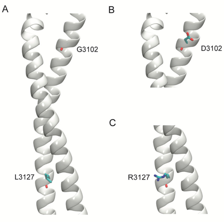
Figure 4.
Models of the stalk region of the wild-type (WT) and variant structures of DNAH11. The stalk region of the protein is represented as a white helix and amino acids are shown in stick representation. (A) WT structure with G3102 and L3127. (B) Structure of the variant G3102D. (C) Structure of the variant L3127R.

