Figure 4.
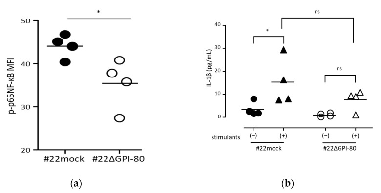
GPI-80 expression augmented NF-κB activation. (a) Measurement of phosphorylated p65-NF-κB levels. The levels of phosphorylated p65-NF-κB in #22mock (closed circle) and #22ΔGPI-80 (open circle) cells were measured by flow cytometry, and the mean fluorescence intensity (MFI) of phosphorylated p65-NF-κB (p-p65 NF-κB) was analyzed. The data represent results from four independent experiments. The statistical significance was calculated using two-tailed unpaired Student’s t-test (*, p < 0.05). (b) Measurement of IL-1β levels. Cells [#22mock (closed symbol) and #22ΔGPI-80 (open symbol)] were incubated with (stimulants (+), triangle symbol) or without (stimulants (−), circle symbol) 10 μg/mL LPS + 1 ng/mL PMA for 24 h. After incubation, the media were collected, and IL-1β concentration in the media was measured. The data represent results from four independent experiments, and the statistical significance was analyzed by one-way ANOVA with Bonferroni’s post hoc test, compared between: #22mock stimulants (−) and #22mock stimulants (+); #22ΔGPI-80 stimulants (−) and #22ΔGPI-80 stimulants (+); and #22mock stimulants (+) and #22ΔGPI-80 stimulants (+) (*, p < 0.05; ns, not significant).

