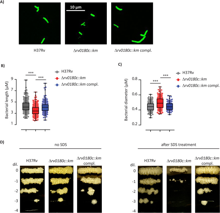
Fig 7. The Δrv0180c::km mutation impacts the bacterial cell shape and cell envelope function.
(A) Fluorescence microscopy image of H37Rv (WT), the Δrv0180c::km mutant, and the Δrv0180c::km complemented strains grown for 10 days in 7H9 ADC liquid medium. (B) Distribution of bacterial length for the parental H37Rv, the Δrv0180c::km mutant and the Δrv0180c::km complemented strains. Between 140 and 220 bacterial cells from two experiments were analyzed. (C) Distribution of bacterial diameter for the parental H37Rv, the Δrv0180c::km mutant and the Δrv0180c::km complemented strains. Around 80 bacterial cells from two independent experiments were analyzed. For (B) and (C), the statistical significance was evaluated using the Kruskal-Wallis rank sum test followed by Dunn’s multiple comparison test: *** p<0.001. (D) Impact of incubation with detergent on the viability of H37Rv, the Δrv0180c::km mutant, and the Δrv0180c::km complemented strains. Bacteria grown in 7H9 ADC to exponential phase (OD600nm between 0.6 and 0.8) were incubated with or without 0.1% SDS during 4 days before spotting 5μl of serial dilution on 7H11 OADC agar plate.

