Figure 2.
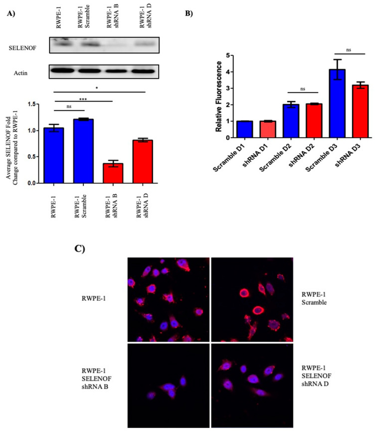
Reduction of SELENOF levels in RWPE-1 cells. (A) SELENOF levels were successfully reduced with an shRNA vector. Western blots using anti-SELENOF antibodies were quantified and a two-tailed t-test was performed for significant differences parental RWPE-1 cells and transfected RWPE-1 cells. n = 3, * p < 0.05, *** p < 0.001. (B) Fluorometric dsDNA quantification was performed after 3 days. Data are represented as the mean ± SEM, ns, not significant, n = 3. (C) Parental RWPE-1 and RWPE-1 scramble cells exhibit SELENOF membrane-associated localization shown in red, similar to what is seen in human benign tissues. Nuclei are stained blue with DAPI. Both shRNA RWPE-1 transfected clones have diffuse SELENOF staining in the cytoplasm similar to what is seen in prostate cancer tissue and prostate cancer cell lines (magnification is 63×).

