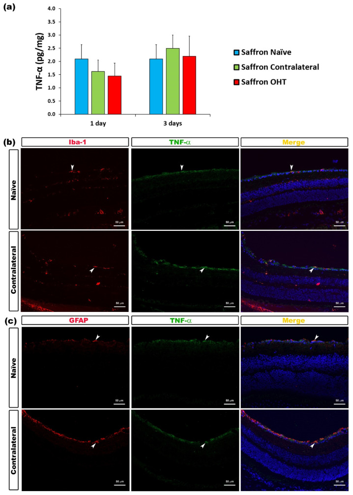
Figure 6.
TNF-α levels at 1 and 3 days after laser-induced ocular hypertension (OHT). (a) The TNF-α values obtained in the multiplex assay. The histogram shows the mean levels (±SD) of TNF-α (pg/mg) at days 1 and 3 after laser OHT induction in saffron ocular hypertension eyes (saffron OHT) and saffron-contralateral eyes (saffron-contralateral) as well as in saffron-naïve eyes. (b) Immunohistochemical study of TNF-α expression in saffron-contralateral eyes three days after unilateral laser-induced OHT. Retinal sections were immunolabeled with antibodies to TNF-α (green) and Iba-1 (red in (b)), or GFAP (red in (c)). Merge is denoted by the colour yellow. (b) The arrowhead shows the co-expression of Iba-1 and TNF-α. (c) The arrowhead shows the co-expression of GFAP and TNF-α. Abbreviations: OHT (ocular hypertension); TNF-α (tumour necrosis factor-α); Iba-1 (ionized calcium-binding adaptor molecule); GFAP (glial fibrillary acidic protein).

