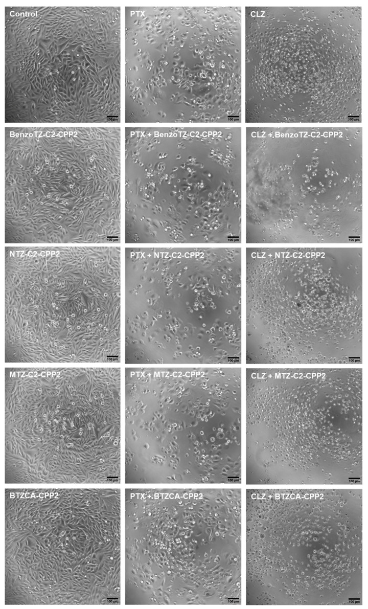
Figure 10.
Microscopic visualisation of PC-3 treated with CPP2-thiazole derivates, both alone and combined with PTX and CLZ, at concentrations of 4 × IV. Representative images were obtained with a high contrast brightfield objective (10×) (LionHeart FX Automated Microscope) from three independent experiments. Scale bar: 100 µM.

