Figure 1.
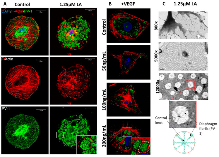
bEND.5 cells have the potential to become fenestrated. (A). BEND.5 cells treated with 1.25 μM of LA had PV-1 (green) and actin (red) redistribution to sieve plates as compared to control non-treated cells (sieve plate indicated by white box and red dotted line). (B). VEGF-induced sieve plate formation could also be stimulated in bEND.5 cells as illustrated by the rearrangement of PV-1 (green) and the actin cytoskeleton (red) (white box and dotted line indicate sieve plate formation in 200 ng/mL of VEGF-treated bEND.5 cells). (C). TEM showed uniform distribution of fenestrae (800× magnification, scale bar: 1 µM; 5000× magnification, scale bar: 0.5 µM). At high magnification, fenestrae diaphragm fibrils and central knots could be visualized (12,000× magnification, scale bar: 200 nm) as highlighted in the accompanying diagram.

