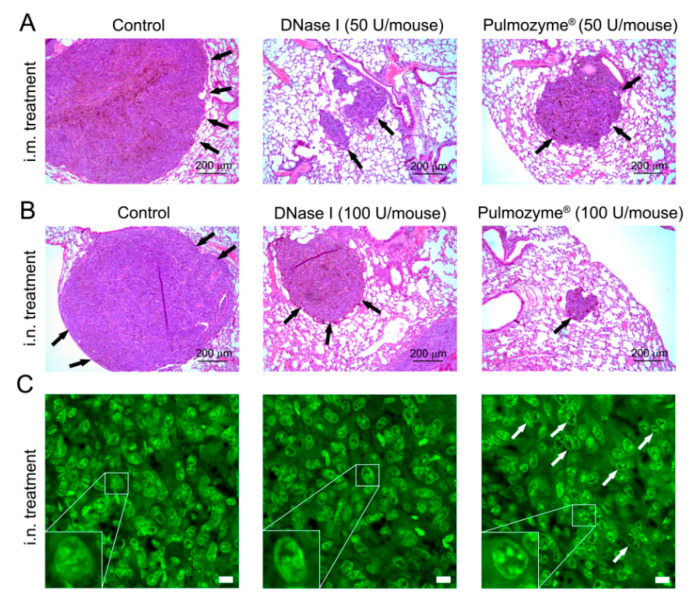
Figure 4.
Representative images of B16 melanoma metastases in the lungs of tumor-bearing mice without treatment (control) and after i.m. and i.n. administration of Pulmozyme® and DNase I. (A,B) Metastases in lungs in intramuscular (A) and intranasal (B) experiments. Hematoxylin and eosin staining, light microscopy, original magnification ×100. Black arrows indicate metastatic foci in the lungs. (C) SYTOTM staining, confocal fluorescent microscopy using a plan-apochromat 63×/1.40 Oil DIC M27 objective. Scale bar corresponds to 10 μm. White arrows indicate B16 melanoma cells with disrupted structural organization of nuclei. Typical examples of B16 cells with condensation and parietal localization of chromatin are shown in the bottom left corner.

