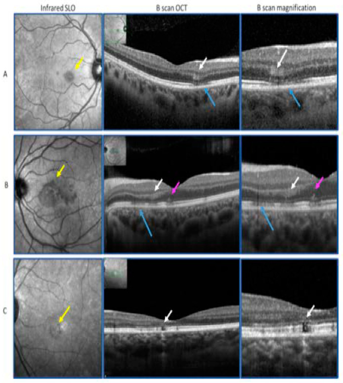
Figure 1.
(A): “Classical” acute macular neuroretinopathy (AMN) with a petaloid, sharp border, lesion on infrared scanning laser ophthalmoscopy (SLO) (yellow arrow), corresponding to an outer plexiform layer (OPL) hyper-reflectivity on optical coherence tomography (OCT) (Heidelberg Engineering, V1.10.12.0, 69115 Heidelberg, Germany) B-scan (white arrows), and with extrafoveal ellipsoid zone (EZ) disruption (blue arrows). (B): AMN “plus” with “photoreceptoritis” with a petaloid, fluffy border, lesion on infrared SLO (yellow arrow) corresponding to OCT B-scan OPL hyper-reflectivity (white arrows), and EZ disruption observed in the foveal center (pink arrows). (C): AMN “plus” with “photoreceptoritis” and “retinal pigment epithelitis”: infrared SLO lesion (yellow arrow) with retinal pigment epithelium (RPE) atrophy on OCT B-scan (white arrows).

