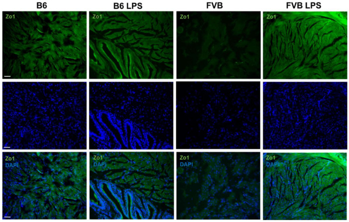
Figure 8.
Immunostaining of zonula occludens-1 (ZO-1, green, first row) for the visualization of tight junction proteins in the bladders of B6-ctrl, B6-LPS, FVB-ctrl, and FVB-LPS mice. DAPI staining and the ZO-1 and DAPI merged images for each group are shown in the second and third row, respectively. All images were acquired at 100× magnification (Scale bar = 50 µm).

