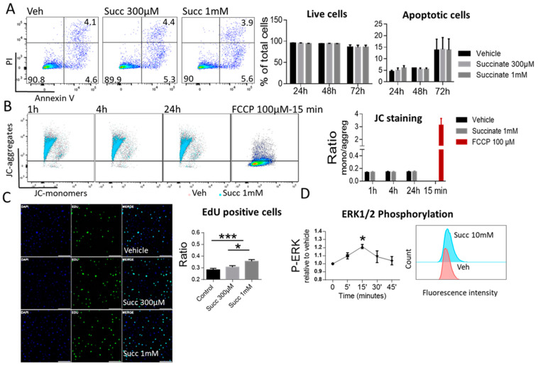
Figure 4.
Succinate Induces proliferation but Not apoptosis of HUVECs. (A) Annexin V/PI staining of HUVECs treated either with vehicle or succinate. The percentages of apoptotic and live cells were calculated at 24, 48 and 72 h. (B) JC-1 staining of HUVECs treated with vehicle or succinate. FCCP, a mitochondrial membrane uncoupler, was used as a positive control. The ratio of JC monomers to aggregates was calculated from median fluorescence intensity at 1, 4 and 24 h. For A and B, representative flow cytometric dot plots are shown, data are mean ±SEM, (n = 3). (C) EdU proliferation assay of HUVECs treated with vehicle or succinate. At least 5 different fields per slide were examined and the ratio of EdU positive cells to the total number per field was calculated. Representative image is included (scale bar 200 µm). (D) ERK1/2 phosphorylation in EA.hy926 cells in response to succinate. Data are expressed as fold change in fluorescence intensity relative to vehicle. Representative histogram is shown. (C,D) were analyzed with one-way ANOVA followed by Tukey’s post hoc test, * p < 0.05, *** p < 0.001. Data are shown as mean ±SEM (n = 3-5).

