. 2021 Oct 22;10:e72171. doi: 10.7554/eLife.72171

Figure 1. Extracellular propionate and butyrate induce histone hyperacetylation.

(A) Histone proteomics of HCT116 cells treated with 1 mM acetate, propionate, or butyrate for 1 hr. (B) Histone acylation over a time course of 1 mM acetate, propionate, or butyrate treatment. (C) SCFA treatment (1 mM, 1 hr). of HEK293, MCF7, and HepG2 cell lines. All values are log2(fold change) over untreated, time-matched controls. *=p≤0.05, **=p≤0.01, ***=p≤0.001. n=3 per condition.

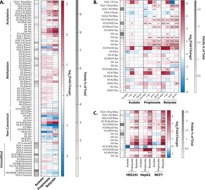
Figure 1.

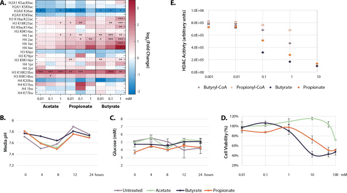
Figure 1—figure supplement 1. Physiological effects of SCFA treatment.

Figure 1—figure supplement 1.

(A) Histone proteomics of HCT116 cells treated with 0.01, 0.1, or 1 mM acetate, propionate, or butyrate for 1 hr. Values are log2(fold change) over untreated control. *=p≤0.05, **=p≤0.01, ***=p≤0.001. (B) Media pH of HCT116 cells over 24 SCFA treatment. (C) Media glucose concentration over 24 hr SCFA treatment. (D) Cytotoxicity curves for acetate, propionate, and butyrate after 24 hr treatment. (E) Values used for HDAC inhibition calculations in Table 1. All experiments were repeated at least two times with an n=3 per condition, representative data is shown. Raw values for B-E can be found in Source data 1. HDAC, histone deacetylase; SCFA, short-chain fatty acid.