. 2021 Oct 22;10:e72171. doi: 10.7554/eLife.72171

Figure 4. p300 inhibition, but not HDAC inhibition, reverses SCFA-induced hyperacetylation.

(A) Acetylation of H3K27ac, H3K18ac, and p53K382ac after treatment with A485, SAHA, and 100–250 µM of propionate/butyrate for 24 hr. Values are normalized to total H3 or total p53 before calculating fold changes to the appropriate untreated control. (B) Acetylation of H3K27ac and H3K18ac in cells transfected with sham, WT p300, or ΔGlu p300 plasmids. Values are fold change over untreated cells with the same transfection. (C) Activity of immunoprecipitated p300 after treatment with 500 µM of propionate or butyrate. Activity is measured with a radioactive assay and normalized to concentration of immunoprecipitated p300 in each sample. *=p≤0.05, **=p≤0.01, ***=p≤0.001. n≥3 per condition. HDAC, histone deacetylase; SCFA, short-chain fatty acid.

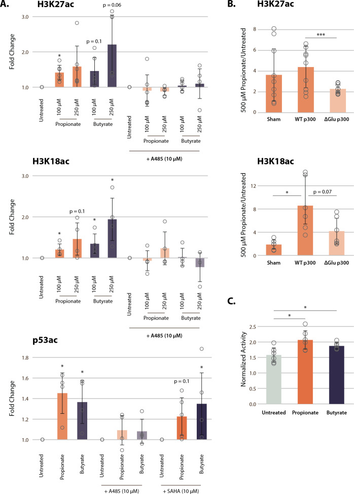
Figure 4.

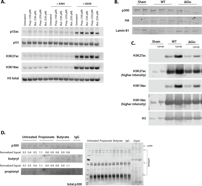
Figure 4—figure supplement 1. Representative Western blots corresponding to Figure 4.

Figure 4—figure supplement 1.

(A) Acetylation of H3K27ac, H3K18ac, and p53K382ac after treatment with A485, SAHA, and 100–250 µM of propionate/butyrate for 24 hr. (B) Expression of HA-tagged plasmids and p300 levels in cells transfected with sham, WT p300, or ΔGlu p300. (C) Levels of H3K27ac, H3K18ac, and H3 in sham, WT p300, or ΔGlu p300-transfected cells. (D) Levels of p300, butyrylation, and propionylation in p300 immunoprecipitated from HCT116 cells treated with 500 µM propionate or butyrate for 1 hr. All experiments were performed at least two separate times with n=3 biological replicates. Values and image quantification for all replicates available in Source data 1, full blots can be found in Source data 2.