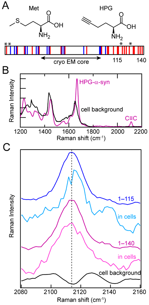
Figure 3.

Biosynthetic incorporation of HPG, a Met analogue, into α-syn as site-specific Raman probes. (A) Structures of Met and HPG. HPG is readily incorporated into α-syn by Met-auxotrophic E. coli. Schematic of α-syn sequence showing acidic (red) and basic (blue) residues, as well as the fibrillar core determined by cryo-EM and the locations of Met residues replaced with HPG (asterisks). The C-terminal truncation site (115) is also indicated. (B) In the Raman spectrum, the alkyne stretching mode (C≡C) of HPG-α-syn (purple) appears in the quiet region of the cellular background (black). (C) The alkyne stretching band of HPG-α-syn1–115 in cells (cyan) is narrower and blue-shifted relative to full-length HPG-α-syn1–140 fibrils in vitro (purple) and in cells (magenta) as well as HPG-α-syn1–115 in vitro (blue).The cell background (black) is shown for comparison. Data originally published in [27].
