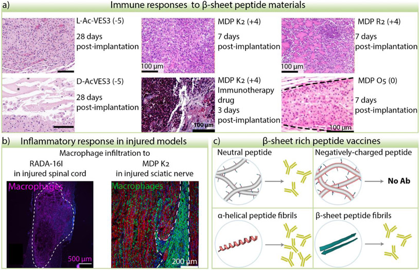
Figure 4.
Immune and inflammatory responses to β-sheet peptide materials. a) Examples of peptide hydrogel sections from subcutaneous injection of materials stained with Hematoxylin and Eosin. Cell nuclei are stained purple, cell cytoplasm, collagen, and hydrogel are stained in different shades of pink. Distinct cell morphology, infiltration degree, and cell distribution is observed depending on the peptide sequence, stereochemistry, immunotherapy drug cargo, and time. L- and D- AcVES3 images were adapted from [100] with permission from the American Chemical Society. Images of MDP K2 and R2 were adapted from [122] and MDP K2 with immunotherapy drug from [123]. © 2020 and 2019 Elsevier Ltd. All rights reserved. Image from MDP O5 was adapted from [106]. © 2019, American Chemical Society. The peptide charge at pH 7 is indicated in the parenthesis. b) When peptide hydrogels are implanted in injured nerve tissue such as spinal cord or sciatic nerve, macrophage infiltration is observed. RADA16-I image adapted from [21]. © 2020 Acta Materialia Inc. Published by Elsevier Ltd. All rights reserved. MDP K2 nerve image was adapted from [109]. © 2020 Elsevier Ltd. All rights reserved. c) Effects of peptide charge and structure in the efficacy of antibody production of epitope-bearing peptide fibers.

