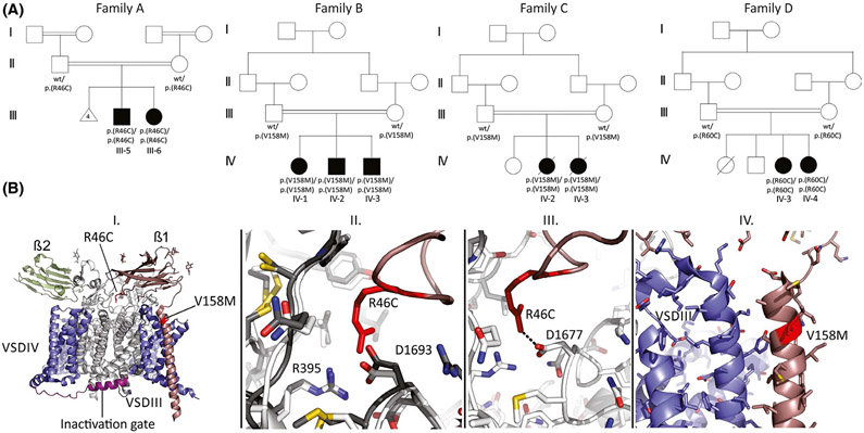
FIGURE 1.
Pedigrees of the studied families and SCN1B variant modeling. (A) multigeneration pedigrees of families A–D showing parental consanguinity and history of recurrent Family A miscarriages. (B) I. Three-dimensional model of NaV1.7 channel encompassing β1 (brown) and β2 (green) subunits. The location of the R46C and V158M mutations in SCN1B (NM_001037.5) is indicated. II. Superposition of NaV1.2 (dark) and NaV1.7 (light) at the contact area for Arg46 in β1, showing that the interface is conserved. Arg46 makes multiple contacts with the channel. III. Dotted line represents an ionic interaction between Arg46 in β1 and Asp1677 in NaV1.7 (corresponding to Asp1693 in NaV1.2). IV. Structural details around the β1 residue V158 are shown

