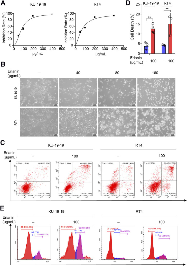
FIGURE 1.
Erianin inhibited cell proliferation and triggered cell death in bladder cancer cells. (A) Cell proliferation of KU-19–19 and RT4 was assessed by CCK-8 assay after the treatment with different dose of erianin treatment for 24 h. (B) The cell morphology was observed by microscope. (C,D) Flow cytometry analysis of cell death by Annexin V-FITC/PI staining in KU-19–19 and RT4 cells were treated with erianin or DMSO control for 24 h, and the quantification of percentage of the cell death was shown. *p < 0.05,**p < 0.01. (E) The percentage of cells treated with erianin in each phase was assessed by flow cytometry.

