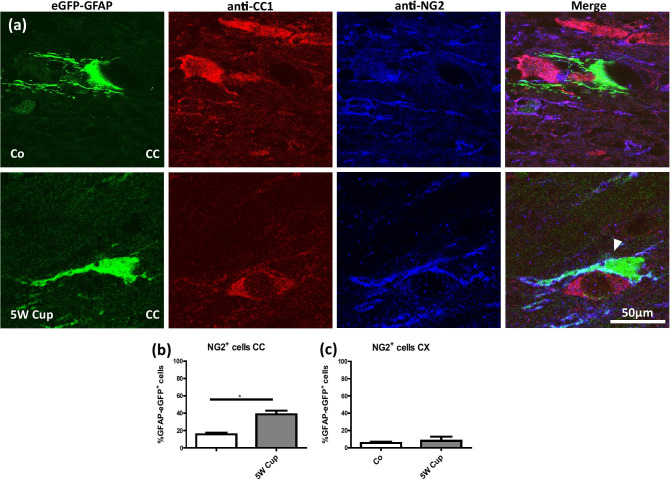
Fig. 4.
Immunohistochemical analysis of anti-CC1 (red), and anti-NG2 (blue) in eGFP-GFAP (green) mice after cuprizone intoxication. (a) Representative sections of eGFP-GFAP, anti-CC1, and anti-NG2 stains in the CC of control (Co) and 5-week cuprizone (5 W Cup) intoxicated mice. The white arrowhead highlights the co-localization of eGFP-GFAP and anti-NG2+. (b) (c) The quantification of eGFP-GFAP/anti-NG2 co-localization in the CC and CX, respectively. Four animals per groups; two independent experiments. Statistically significant differences are indicated by asterisks (*p < 0.05). Scale bar = 50 µm

