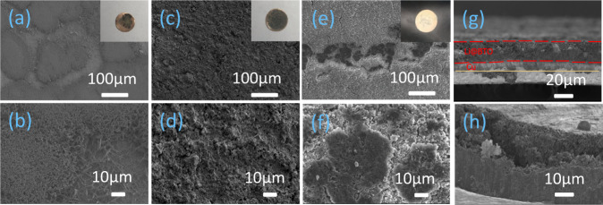
Fig. 7. Ex situ SEM postmortem morphological investigations of the bare Cu, AO and BTO scaffold after plating to 1 mA h cm−2 at a current density of 2 mA cm−2.
a, b Bare copper electrode and zoomed-in figure, c, d BTO scaffold and zoomed-in figure, e, f AO scaffold and zoomed-in figure, g, h cross-section of BTO and Cu electrode after depositing 1 mA h cm−2 Li metal at a current density of 2 mA cm−2. Insets show the digital images of the complete electrodes.

