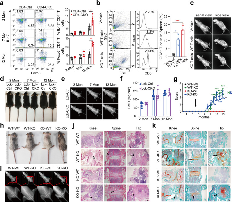
Fig. 3. Immune system abnormality is not the cause of AS-like bone deformation.
a Flow cytometry analysis of T cell subsets in inguinal lymph nodes. (n = 5). b Flow cytometry results of CD3+ T percentages in the spleen of 6-month-old recipient NCG mice (n = 4). c X‐ray images of 12-month-old recipient NCG. d–f Gross images (d), X‐ray images (e), and Femoral BMD (f) of Lck-CKO mice and Lck-Ctrl littermates. (n = 5). g–k CD4-Ctrl (WT) and CD4-CKO (KO) mice at 4 months were lethally irradiated followed by transferring with bone marrow cells. Irradiated CD4-CKO mice transferred with WT bone marrow cells were labeled as WT-KO, and so on. Pathological scores (n = 10) (g), Gross images (h) and Radiographs (i) of 12-month-old chimeras. c–e, h–i Scale bars: 1 cm. j, k H&E and SOFG staining images of chimeras. Scale bars: 200 μm. j Arrows show bone deformation. k Arrows indicate chondrogenesis and ectopic new bone formation. a, b, f, g Data are presented as mean ± SEM. *p < 0.05. **p < 0.01, ***p < 0.001, determined by two-tailed Student’s t-test. c–e, h–k Data are representative of three independent biological replicates.

