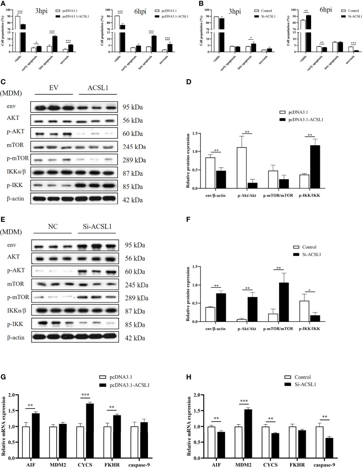
Figure 4.
ACSL1 induced apoptosis via PI3K/Akt signaling pathway. (A, B) ACSL1 overexpression (A) and knockdown (B) in MDMs for 48 h, followed by infection with ALV-J (104 TCID50/0.1 ml) before assays. Apoptosis analysis of MDMs for 3 and 6 hpi, using Annexin V-FITC. Statistical analysis of the data from the multiple repeated Annexin V-FITC experiments. (C, D) Immunoblot analysis of the levels of env, Akt, p-Akt, mTOR, p-mTOR, IKKα/β, and p-IKK expression in MDMs transfected with ACSL1 expression plasmids or empty vector control for 48 h followed by ALV-J infection for 3 h. (E, F) Immunoblot analysis of the levels of env, Akt, p-Akt, mTOR, p-mTOR, IKKα/β, and p-IKK expression in MDMs transfected with siACSL1 or control siRNA for 48 h followed by ALV-J infection for 3 h. (G, H) qRT-PCR analysis of apoptosis-related genes (AIF, CYCS, FKHR, MDM2, and caspase-9) in ACSL1 overexpressed cells (G) or ACSL1 knockdown cells (H) followed by ALV-J infection for 3 h. Data shown are the means ± SEM (n=3). P values were calculated using two-tailed unpaired Student’s t-test. Differences with P < 0.05 were considered significant. *P < 0.05, **P < 0.01, ***P < 0.001.

