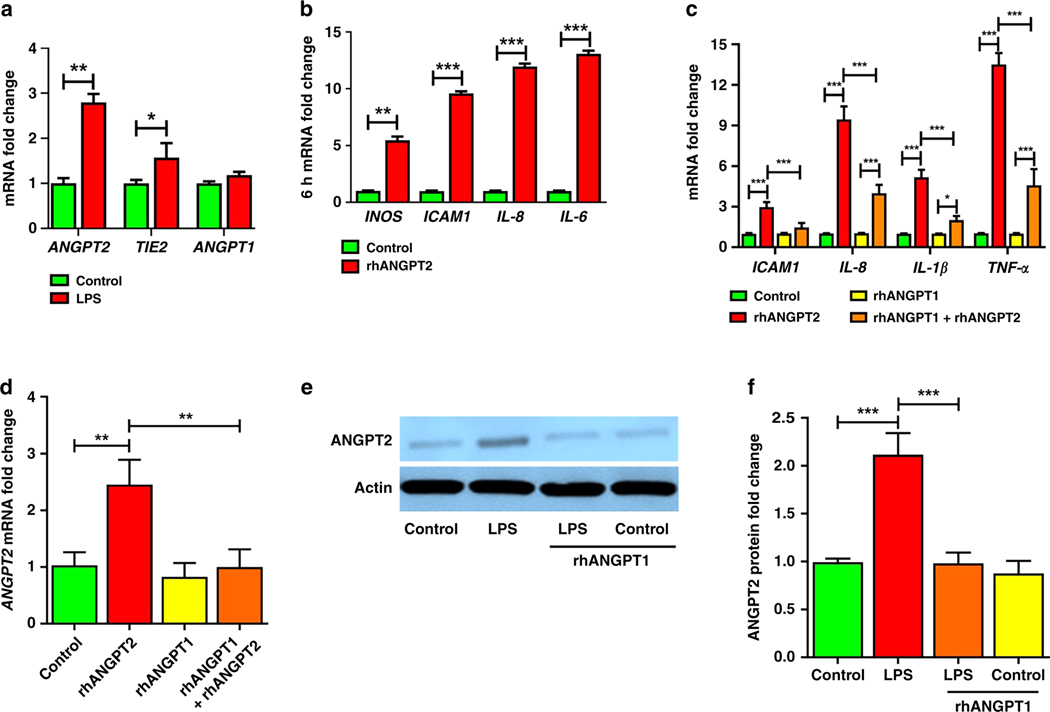
Figure 2– ANGPT2 is upregulated by LPS in HPMEC and directly mediates EC immune activation, which is attenuated by rhANGPT1.
HPMEC were treated with LPS (100ng/mL), recombinant human ANGPT2 (rhANGPT2, 25ng/mL), and rhANGPT1 (200ng/mL), with lysates used for assays. (A) ANGPT1, ANGPT2, and TIE2 gene expression by qPCR 24h after LPS. (n=4/condition, *p<0.05, **p<0.01.) (B) Quantification of INOS, ICAM1, IL-8, and IL-6 expression by qPCR 6h after rhANGPT2. (n=4/condition, **p<0.01, ***p<0.001.) (C-D) HPMEC were treated for 24h with rhANGPT1 and rhANGPT2, and lysates were used to quantify gene expression of IL-1β, ICAM1, IL-8, and TNF-α (C) and ANGPT2 (D) by qRT-PCR. (n=3/condition, *p<0.05, **p<0.01, ***p<0.001 between Control and rhANGPT2; rhANGPT2 and rhANGPT1+rhANGPT2; rhANGPT1 and rhANGPT1+rhANGPT2.) (E) ANGPT2 protein was quantified by immunoblot of HPMEC lysates 24h after rhANGPT1 and rhANGPT2 treatments, with densitometry shown (F). (n=4/condition, ***p<0.001 between Control and LPS; LPS and LPS+rhANGPT1). ANOVA (with Tukey) or Mann Whitney tests were used.

