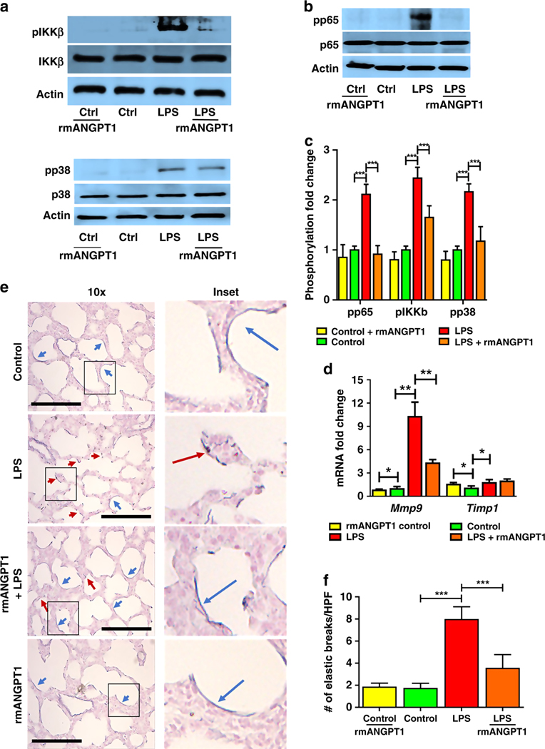
Figure 5– rmANGPT1 modifies TLR4 signaling and matrix degradation during sepsis in mice.
6-day-old pups were injected with LPS and rmANGPT1 pre-treatment, and lungs were harvested 24h post-treatment for assays and staining. (A-C) Lung homogenates were used to quantify the phosphorylation of IKKb and p38 (A), and p65 (B) by immunoblotting, with densitometry shown (C). (n=5/condition, ***p<0.001 between Control and LPS; LPS and LPS+rmANGPT1.) (D) Mmp9 and Timp1 gene expression by qPCR from lung homogenates. (n=5/condition, *p<0.05, **p<0.01 between Control and LPS; LPS and LPS+rmANGPT1; Control and Control+rmANGPT1.) (E) Lung tissue sections were assessed for elastic fiber structure (blue) using Resorcin-Fuchsin staining with Carmine counter stain, with disrupted elastic fibers shown by red arrows and continuous fibers shown by blue arrows. Scale bar indicates 30μm. (F) Quantification of elastic fiber breaks/HPF, is shown. (n=5/condition, ***p<0.001 between Control and LPS; LPS and LPS+rmANGPT1.) ANOVA (post-hoc Tukey tests) or Mann Whitney tests were used.

