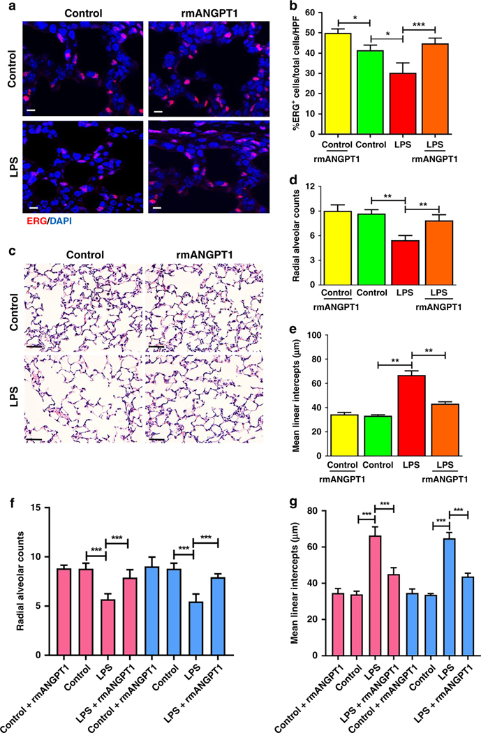
Figure 6– Effect of rmANGPT1 on LPS-induced long-term lung growth and remodeling in mice.
Sections of formalin-inflated lungs from 15-day-old mice, 196h post-LPS treatment, with or without rmANGT1 treatments (one dose 2h prior to LPS and another 72h after LPS) were stained. (A) Immunofluorescent staining of ERG (red) and DAPI (blue), with quantification of ERG+/Total cells/HPF shown (B). Scale bar indicates 10μm. (n≥5/condition, *p<0.05, ***p<0.001 between Control and LPS; LPS and LPS+rmANGPT1; Control and Control+rmANGPT1.) (C-E) H&E staining (C) used to quantify radial alveolar counts (D) and mean linear intercepts (E). Scale bar indicates 50μm. (n≥5/condition, **p<0.01 between Control and LPS; LPS and LPS+rmANGPT1.) (F) and (G) Radial alveolar counts and mean linear intercepts were further analyzed by mouse sex. Pink bars represent females and blue bars represent males. (n≥3/condition, ***p<0.001 between Control and LPS; LPS and LPS+rmANGPT1). ANOVA with Tukey tests were used.

