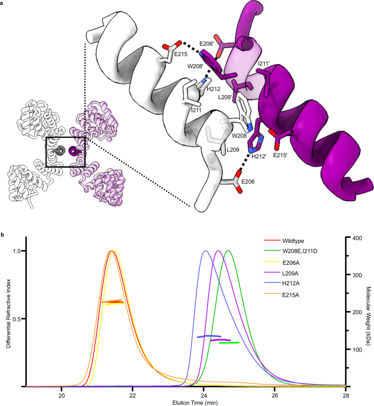
Fig. 2. Probing the dimeric state.
a The crystal structure of the 11.5 TPR construct with helices presented in expanded view depicted in darker shades. Enlarged view with residues subject to mutagenesis depicted. Dashed lines indicate hydrogen bonding. Purple and white denote separate chains, which form the dimer b SEC-MALLS trace demonstrating the multimeric state of OGT. Differential refractive index value depicted as a single continuous trace across the X-axis, while molecular weight estimates shown just for the region analysed. Source data are provided as a Source Data file.

