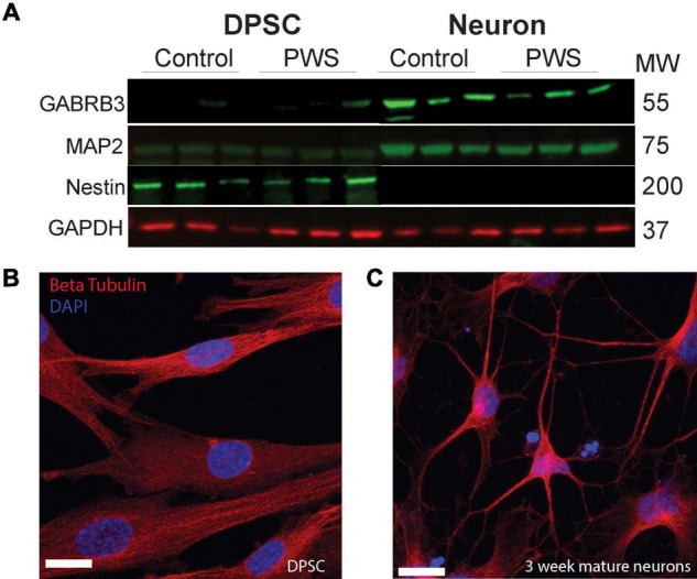
FIGURE 2.

DPSC from both PWS and control subjects differentiate efficiently into neuronal cultures. (A) DPSC from all subjects are negative for neuronal markers, MAP2 and GABA A, but positive for stem cell marker NESTIN. After differentiation and 3 weeks of maturation, the cultures are positive for both MAP2 and GABA A receptor subunit beta 3. (B,C) Representative DPSC versus DPSC-derived neurons visualized with anti-beta tubulin (red) (B) DPSC show a flat fibroblast-like morphology as previously reported. (C) DPSC-derived neuronal culture contains cells with pyramidal neuron morphology. DAPI (blue) was used as a nuclear stain. 63× magnification. Scale bar is 20 μM.
