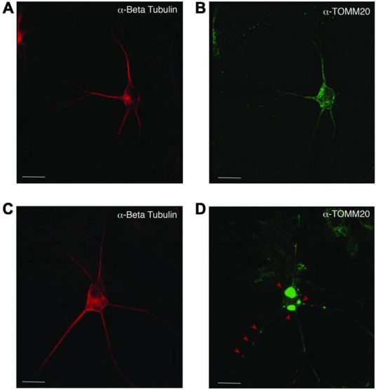
FIGURE 5.

Mislocalization and reduced dispersion of mitochondria in ASD neurons. Neuronal cultures visualized with anti-beta tubulin (red) and the mitochondrial marker anti-TOMM20 (green). The PW-UPD -ASD neuron (top row) shows bright and evenly dispersed mitochondria within the neuronal projections. In PW-UPD + ASD neuron, the red arrows point to mitochondrial aggregates not seen in the PW-UPD -ASD neurons. (A,C) show neuronal morphology using anti-beta tubulin staining. (B,D) show the anti-TOMM20 staining to identify mitochondria. Confocal stacks were taken at 63X magnification. Scale bar is 20 μm.
