
FIGURE 7.
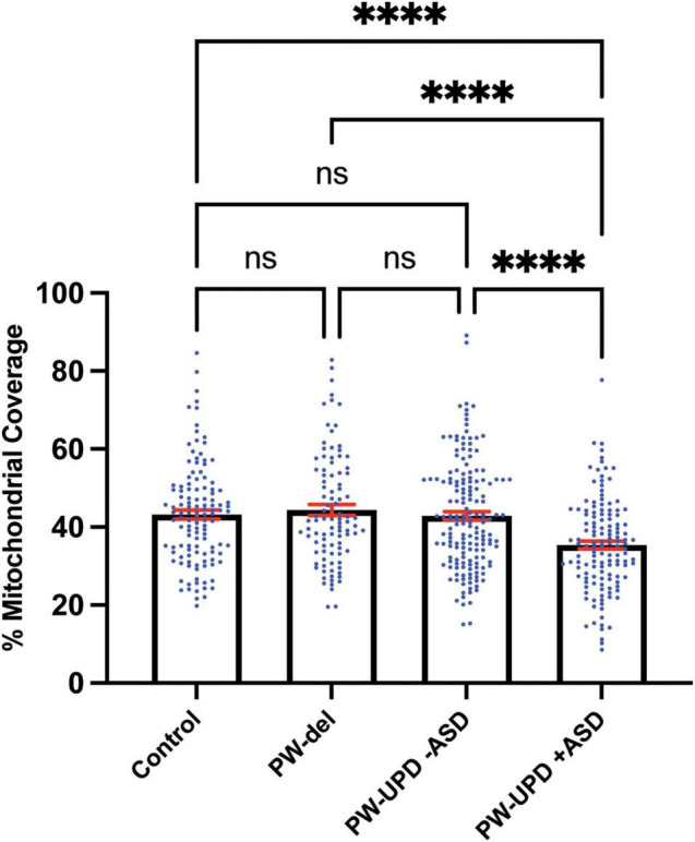
PW-UPD + ASD neurons show decreased mitochondrial coverage within neuronal area. Using Imaris, the total neuronal area (anti-Beta Tubulin) that contains mitochondria (anti-TOMM20) was calculated. Percent of mitochondrial coverage was measured for ≥15 neurons from each cell line and at least 5 cell lines per group in a blinded fashion. Significance testing was performed by both ordinary one-way ANOVA and Turkey’s multiple comparisons test with a single pooled variance. ns = not significant, **** = p-value ≤ 0.001.