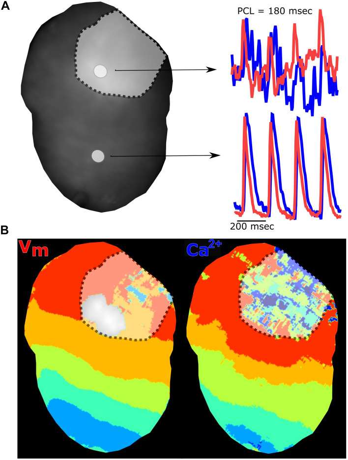
FIGURE 9.
Myocardial injury via radiofrequency ablation diminishes electrical and calcium signals. (A) Langendorff-perfused rat heart after radiofrequency ablation showing two regions of interest and corresponding traces. The voltage signal (red) and calcium signal (blue) are collected from the ablation site (denoted with dotted line) and non-ablated tissue. (B) Voltage and calcium isochronal maps display activation time across the heart, with disruption at the site of ablation injury. Epicardial dynamic pacing from the apex of the heart.

