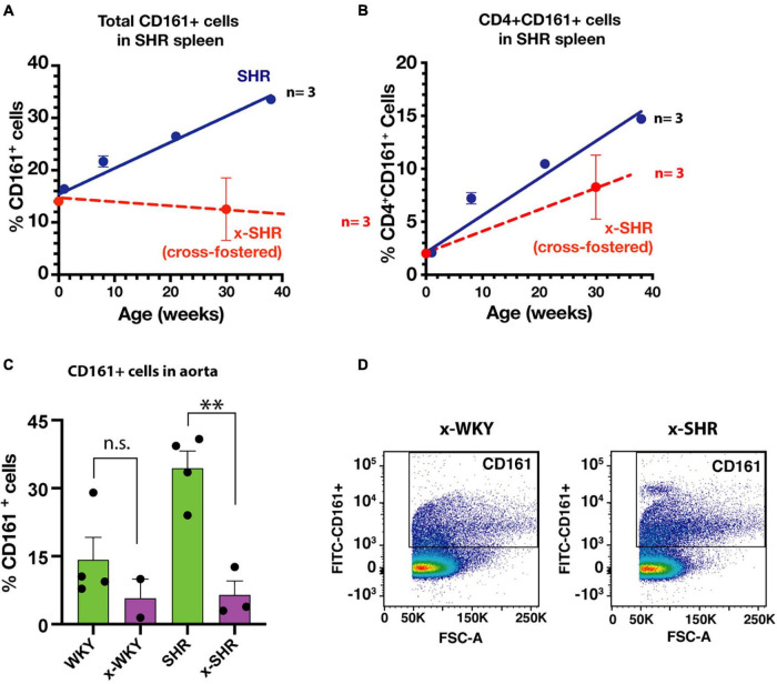
FIGURE 4.
(A) Cross-fostering of SHR prevents the age dependent increase in CD161 + cells and (B) CD4 + CD161 + cells observed in self-fostered SHR as previously reported (Singh et al., 2017) (Blue line). (C) Comparison of infiltration of proinflammatory CD161 + immune cells in aortic tissue in self-fostered cross-fostered WKY and SHR. (D) Representative dot plot of CD161 + cells in thoracic aorta of cross-fostered WKY (x-WKY) and cross-fostered SHR (x-SHR). Asterisks denote statistically significant (∗∗P < 0.0075), whereas n.s. denotes statistically not significant results by one-way ANOVA and Sidak’s post hoc test.

