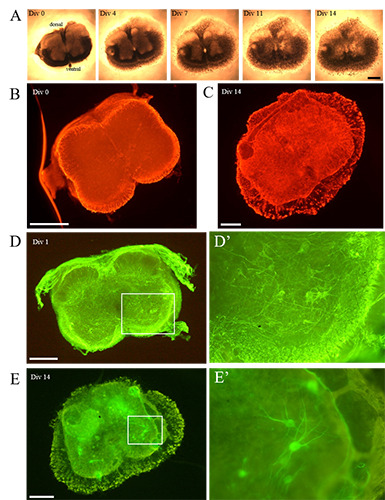
Figure 1.

A) The time lapse analysis shows the gradual modifications occurring to a representative organotypic spinal cord SMA slices, during the 14 DIV: although a progressive thinning of the section was evident (from 4 DIV), overall the slice morphology appeared well preserved during the whole period of observation. B) Immediately after the tissue chopper cutting, no signs of astrogliosis (GFAP) were evident (here showed a SMA slice), whereas C) it increased over time (as visible in this representative WT slice). D) Concerning the cell preservation, at 1 DIV the WT MNs (SMI32- positive cells) were in good conditions and showed numerous neurites (well visible in the inset D’). E) At 14 DIV, the WT MN survival was still good: the cells were correctly located in the ventral horns and several extensions were detectable (a detail is appreciable in the inset E’). Scale bar: A-E) 500 μm; D’, E’) 250 μm.
