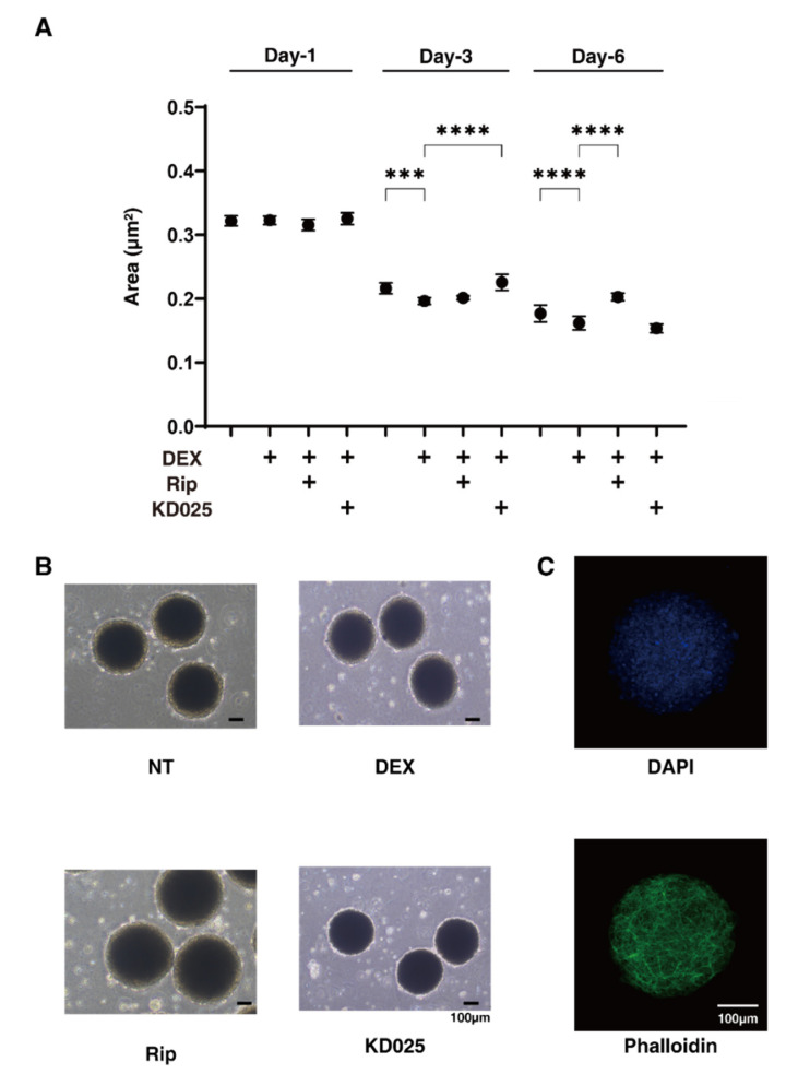
Figure 2.
Changes in DEX-treated 3D HTM spheroid size at Days 1, 3, or 6 in the presence and absence of ripasudil or KD025, and a representative non-treated 3D spheroid image stained with DAPI and phalloidin (C). At Days 1, 3, or 6, the mean sizes of HTM 3D spheroids (non-treated control; NT) and those treated with 250 nM DEX were plotted in the absence or presence of 10 µM ripasudil (Rip) or KD025 (A). (B) shows representative phase-contrast microscope images of the 3D HTM spheroids at Day 6 under several conditions. (C) shows a representative image immune-stained with DAPI and phalloidin of non-treated 3D HTM spheroid. These experiments were performed in triplicate using fresh preparations (n = 10–15). Data are presented as the arithmetic mean ± standard error of the mean (SEM). *** p < 0.005, **** p < 0.001 (ANOVA followed by a Tukey’s multiple comparison test). Scale bar; 100 µm.

