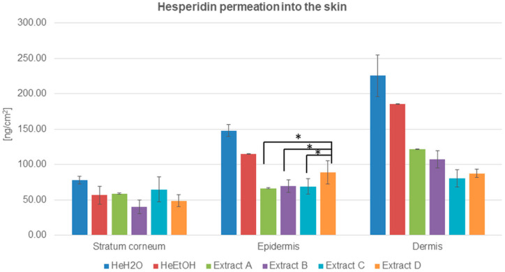
Figure 1.
Hesperidin distribution among skin layers: stratum corneum, epidermis, and dermis (ng/cm2) after application and 24 h of incubation with hesperidin solutions: water (HeH2O, 4 μg/mL), ethanol (HeEtOH, 4 μg/mL, 50% (v/v)), or honeybush extracts: A, B, C, D. Each average value was obtained from three independent repetitions. Error bars represent standard deviations. Significant differences among samples are marked with an asterisk (p < 0.05).

