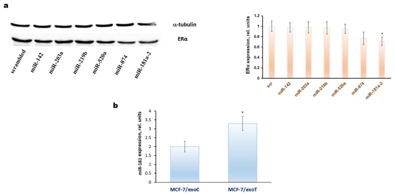
Figure 1.
(a) Influence of miRNAs transfection on the ERα expression in the MCF7 cells. The single transfection by the miRNAs mimetics was performed as described in Methods. Twenty-four hours after transfection the Western blot analysis of ERα expression in the cell lysates was performed. Protein loading was controlled by membrane hybridization with α-tubulin antibodies. Densitometry for immunoblotting data (right diagram) was carried out using ImageJ software (Wayne Rasband, NIH) with the recommendations from the work [83]; * p < 0.05 versus scrambled (scr); (b) quantification of endogenous miR-181a expression (vertical axis) in the exosome-treated MCF-7 cells by qRT-PCR. The MCF-7 cells were cultured in the presence of the exosomes isolated from MCF-7 (exoC) and MCF-7/T (exoT) cells for 30 days with following cell cultivation within the next 30 days after exosomes withdrawal. Three separate measurements were performed for each sample. The expression of RNU6B was used as an internal control. Error bars indicate standard deviation; * p < 0.05 versus MCF-7 cells treated with exoC.

