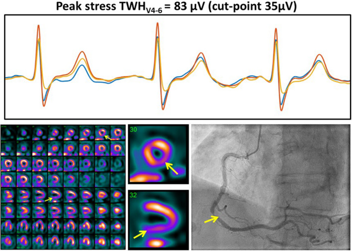
FIGURE 9.

True‐positive myocardial perfusion imaging (MPI) case confirmed by coronary angiogram showed a sizeable increase in TWHV4–6. Regadenoson testing elicited a 30% increase in TWHV4–6 to 83 µV in an 81‐year‐old woman with chest pain (upper panel). Positive MPI revealed a significant lesion in the right coronary artery territory (lower left panel). Coronary angiogram confirmed an 80% lesion in the right coronary artery (lower right panel). The clinical notes indicated reversible, medium‐sized, moderate severity perfusion defect involving the LAD territory; transient cavity dilation consistent with multi‐vessel or left main disease; and normal left ventricular cavity size and systolic function at rest with stress‐induced global hypokinesis. Reprinted with permission from Araujo Silva et al. (2020)
