
Fig. 8.
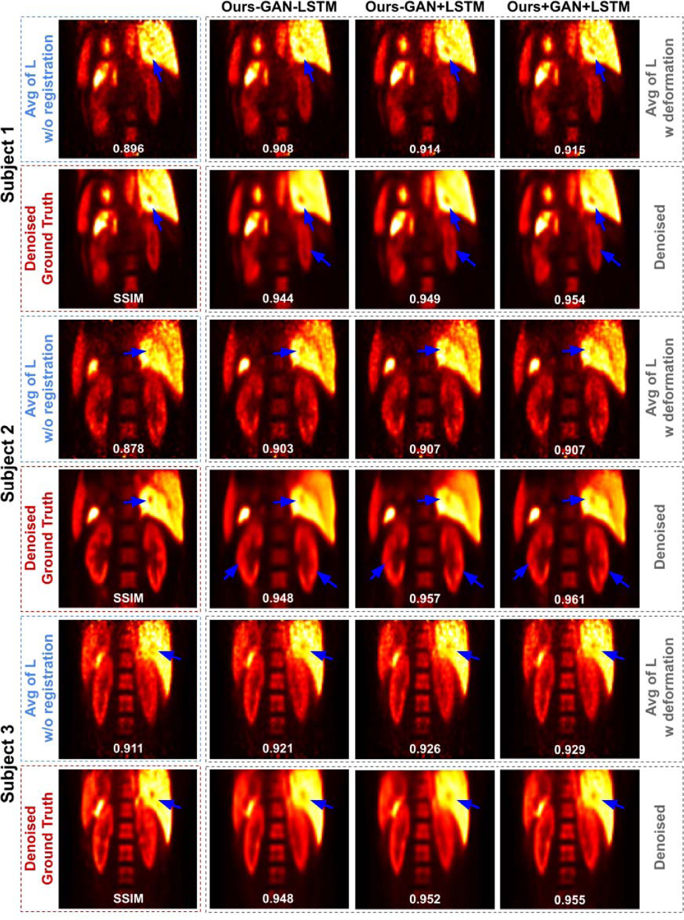
Three subjects with low-dose gated PET. The averaged images L and the corresponding denoised image from different MDPET configurations are shown in the 1st row and 2rd row in each patient’s image group. Motion blurred anatomic structure are recovered using our MDPET (blue arrows).