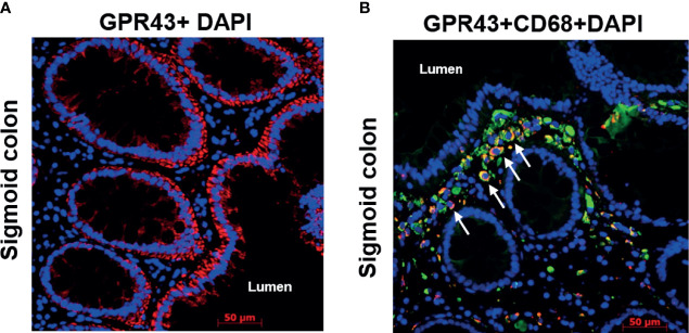
Figure 5.
Immunofluorescence of GPR43 of a representative patient biopsy. Time from transplant to biopsy: 3.5 years, no GvHD at the time of biopsy. (A) GPR43 staining in the sigmoid colon of a patient. GPR43 is labelled with AlexaFlour (AF) 594 (red). (B) GPR43 and CD68 co-staining in the sigmoid colon of a patient. GPR43 is labelled with AF594 (red) and CD68 is labelled with AF488 (green). White arrow indicates colocalized signals. Nucleus is counterstained with DAPI (blue). Scale bar: 50 µm.

